Intervertebral disc disease (IVDD) is a common condition in certain dog breeds that can damage the spinal cord, resulting in significant pain and impaired mobility. Our Countryside Veterinary Hospital team wants to provide information about this disease, in case your dog is at risk.
Intervertebral disc disease in dogs
Numerous small bones (i.e., vertebrae) compose the backbone, and the spinal cord runs through a protected canal in the center of the vertebrae. The vertebrae are separated by spongy intervertebral discs that act as shock absorbers to mitigate concussive forces placed on the spinal cord, and they minimize compressive effects and damage from aging and vigorous activity. Each disc is made of a fibrous outer shell (i.e., annulus fibrosus), a jelly-like interior (i.e., nucleus pulposus), and cartilage caps on each side that connect the structure to the vertebral bones. Changes in the intervertebral disc can lead to IVDD. Two disease processes can affect the disc, including:
- Type 1 — The intervertebral disc’s nucleus pulposus calcifies and extrudes through the annulus fibrosus. The disc material can extrude upward, putting pressure on the ligament that runs above the spinal cord, or sideways, putting pressure on nerves as they exit the spinal cord. Chondrodystrophoid breeds, such as dachshunds, beagles, and Pekingese, are at higher risk. Signs typically present in these dogs between 3 and 6 years of age.
- Type 2 — Type two is a slow degenerative process. The annulus fibrosus collapses, and the outer and inner disc portions place pressure on the spinal cord. German shepherds and Labrador retrievers are at higher risk, and signs typically present between 5 and 12 years of age.
Intervertebral disc disease clinical signs in dogs
IVDD signs depend on the disc herniation location.
- Cervical IVDD — Disc herniations in the neck region typically affect chondrodystrophoid breeds, with neck pain the predominant sign. The dog may have difficulty holding up their head, and will commonly hold their head low and straight out, and vocalize in pain if their head is manipulated. In severe cases, forelimb lameness may be present.
- Thoracolumbar IVDD — Space in the vertebral column is smaller in the back and pelvis region, so less herniated material is needed to cause clinical signs. Dogs will experience pain over the affected area, and may also exhibit incoordination, hindlimb paresis or paralysis, and urinary or fecal incontinence.
Intervertebral disc disease diagnosis in dogs
When a dog presents with spinal pain and weakness, our veterinary professionals must determine if the spinal cord is compressed. Diagnostics include:
- Neurological examination — We assess your dog’s ability to move, palpate their neck and back, and test different reflexes to determine where the spinal cord is affected. We also perform tests to determine if your dog has lost pain perception.
- X-rays — Regular X-rays can rule out issues such as broken bones or dislocations, and they can confirm about 60% of IVDD cases.
- Advanced imaging — Myelography involves injecting a contrast dye around the spinal cord to detect the compressed area. In some cases, an accurate IVDD diagnosis will require computed tomography (CT) or magnetic resonance imaging (MRI).
Intervertebral disc disease conservative treatment in dogs
If your dog can walk and is not in extreme pain, conservative treatment is a reasonable choice. Often the dog is hospitalized for the first week to carefully monitor their progress. Treatments include:
- Confinement — Strict confinement is the most important aspect of conservative IVDD management. The dog should be placed in a cage or crate, and allowed out only for brief bathroom breaks, when they should be carried, if possible. Cage rest for three weeks is a minimum treatment course.
- Medications — Non-steroidal anti-inflammatories, steroids, and muscle relaxants are commonly prescribed to help manage the inflammation and pain.
- Ice packs — The affected area can be ice-packed for 10 to 15 minutes several times a day to help decrease inflammation.
- Physical rehabilitation — Therapeutic exercises are often recommended to improve functional ability.
Intervertebral disc disease surgical treatment in dogs
Spinal surgery is highly invasive, but is necessary in some cases to preserve the dog’s ability to walk. The longer the neurological deficits have been present, the poorer the dog’s prognosis. The best candidates for spinal surgery are dogs who cannot walk but still have deep pain perception in at least one limb. If a dog has been unable to walk for less than 48 hours and has no deep pain perception in their hindlimbs, surgical success drops to 50% to 60%. After 48 hours, the prognosis for recovery is much worse. Post-surgical care, which involves expressing the dog’s bladder, nursing care to prevent urine scald and bed sores, and physical rehabilitation exercises, can be intense. Dogs typically must remain confined for four to six weeks after surgery.
Intervertebral disc disease prevention in dogs

Deterioration leading to IVDD cannot be prevented, but you can take steps to decrease your dog’s risk. These include:
- Keeping your dog at a healthy weight — Overweight dogs are predisposed to IVDD. Calculate your dog’s daily caloric needs and feed them appropriate portions to keep them at a healthy weight.
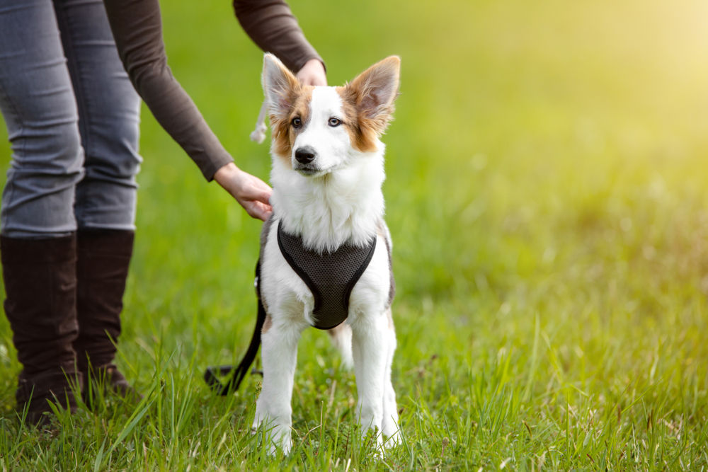
- Using a harness-style collar — When walking your dog, reduce neck strain by using a harness that distributes the weight across their chest and away from their neck.
- Minimizing high-impact activity — In high-risk dogs, minimize high-impact activities such as jumping on and off furniture.
IVDD is a debilitating and painful condition, and affected dogs should be treated on an emergency basis. If your dog is experiencing neck or back pain, contact our Countryside Veterinary Hospital team, so we can alleviate their distress and get them back on their feet as soon as possible.


Leave A Comment